From galaxies to neurons
This story originally appeared in Yale Engineering magazine.
How does a brain form a memory, and why does it store certain ones, while others fade away? How does it allow a person to learn? These questions are at the core of what makes the brain so mysterious. But what if we had the technology to better see the brain’s hardware — the neurons, synapses, and so much else — that would allow us to answer these questions?
That’s what Cristina Rodríguez is working on. A big part of her lab’s mission is combining her backgrounds in physics and engineering to develop optical microscopy tools to better visualize critical biological processes as they happen naturally, with a particular focus on applications in neuroscience. That is, she’s building new and better microscopes to see what couldn’t be seen before.
Read more • Approximately 5 minutes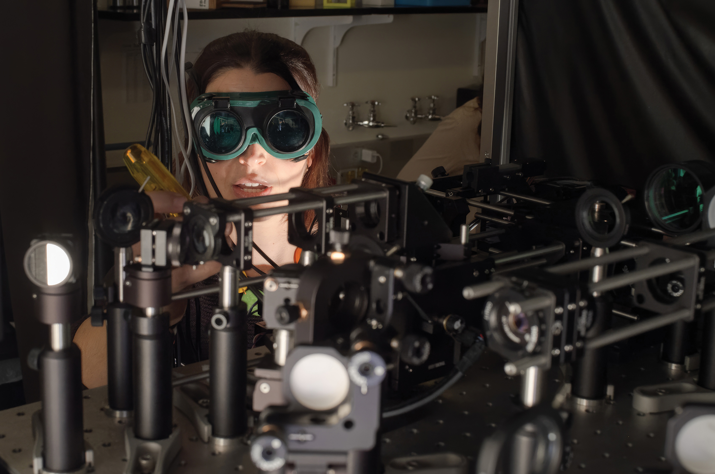
Using cutting-edge imaging techniques, she and her research team were among the first to view with unprecedented detail neurons and calcium signaling in the spinal cord of a mouse. These neurons were detected in tissue three times deeper than had ever been observed before. One key finding was that temperature-sensitive neurons existed in these areas of the spinal cord — something previously unknown.
“If I changed the temperature somewhere in the skin of the mouse — let’s say the hind limb — I could see that there is a neuron firing,” said Rodríguez, assistant professor of biomedical engineering. “And then every time I cool the skin, that neuron fires.”
Rodríguez arrived at this discovery using a combination of technologies: three-photon fluorescence microscopy and adaptive optics. With multiphoton microscopy, tightly focused femtosecond laser pulses at near-infrared wavelengths excite fluorescent molecules, enabling precise high-resolution imaging hundreds of microns deep within tissues. Two-photon fluorescence microscopy, which has been in use for more than three decades, has allowed researchers to observe biological structures and processes deep inside living organisms. Its use is limited, however, especially when imaging deep brain layers. That’s where the more recently developed three-photon fluorescence microscopy comes in. Here, three photons are simultaneously absorbed — instead of two, as in two-photon fluorescence microscopy — allowing the use of longer excitation wavelengths, which reduce light scattering. That provides better penetration into tissues.

Adaptive optics is a technology borrowed from the field of astronomy, where it’s used to correct for aberrations from the atmosphere to get a clearer picture of stars and other celestial bodies. As it turns out, the same technology is as useful for observing neurons as it is for stars.
“It’s a very powerful technology that is also incredibly useful in the context of microscopy,” Rodríguez said. “I’m not trying to look at stars in the center of our galaxy, but synaptic structures deep inside our brain. As part of my work, I’ve shown how we could do this deep inside of a mouse brain.”
Before starting her journey in microscopy development and biological imaging, she spent a few years studying astronomy at The University of New Mexico in Albuquerque, first during a study abroad program as an undergraduate and later as a physics graduate student. Among other findings, she discovered two supermassive black holes at the center of a galaxy, located 24 light years apart — more than 100 times closer than any pair that had been detected previously, offering critical insights into galaxy evolution.
“It was very exciting, but as a physicist, I was really drawn to the instrumentation side,” she said. “I thought it would be really exciting to build an instrument that allows you to see something otherwise invisible.”
“Who’s not interested in the brain, right? It’s so intriguing. I was always reading books about the brain and I found it absolutely fascinating. And particularly, there was something about memories that fascinated me. How do we form memories? How does our brain decide when to forget something? Maybe as a protective mechanism. And why are some memories so vivid and powerful?
Cristina Rodríguez
Assistant Professor of Biomedical Engineering
Specifically, that includes the chance to see those ineffable phenomena produced by the brain. After being drawn by the mysteries of the universe, she then veered in the other direction.
“Who’s not interested in the brain, right? It’s so intriguing,” she said. “I was always reading books about the brain and I found it absolutely fascinating. And particularly, there was something about memories that fascinated me. How do we form memories? How does our brain decide when to forget something? Maybe as a protective mechanism. And why are some memories so vivid and powerful?”
Before biomedical engineering and before astronomy, Rodríguez had her sights on becoming a doctor, like both of her parents while growing up in Venezuela. Her father is a cardiologist, and her mother a hematologist. When she was about 10 years old, she constantly wore a Yale School of Medicine shirt featuring Snoopy in surgical scrubs — a gift from her mother after a conference in New Haven.
“I proudly wore that shirt, hoping that one day I might be at Yale. It probably would have sounded like a crazy dream, but here I am,” she said. “It feels like coming full circle. While I took a different path than becoming an M.D., building microscopes to study biology will allow me to make a meaningful impact in medicine.”
More Details
Published Date
Apr 18, 2025


